Epidermal Grafting in Pyoderma Gangrenosum – JAMA Derm
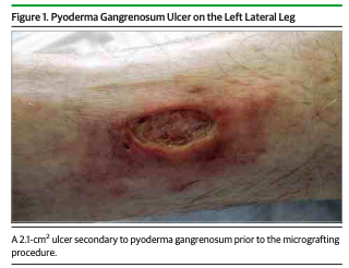
Report of Cases Patient 1 A woman in her 40s without other significant medical history presented in 2013 with an 8-month history of an exquisitely painful ulcer on the distal left lateral leg. She noted that the lesion began as “a small red bump” that she believed to be an insect bite. A physician at another institution performed an incision and drainage to treat a suspected abscess and prescribed oral antibiotics, but the lesion deteriorated. A biopsy performed in 2012 had nonspecific results and Gram, acid-fast, and Fite stains were negative for organisms. Venous and arterial insufficiency were excluded by lower limb duplex ultrasound.She received a diagnosis of pyoderma gangrenosum (PG) and was prescribed prednisone 60 mg daily and topical clobetasol propionate ointment, 0.05%, to the wound edges. On referral, physical examination revealed a sharply demarcated ulcer with purple undermined borders, consistent with the PG diagnosis. The prednisone dosage was tapered because of adverse effects, and cyclosporine 400 mg daily and weekly leg compression were initiated. Although the inflamed undermined borders improved, the wound failed to reduce in size after 2 months of therapy (Figure 1). Patients 2 Through 5 Four additional patients with chronic, recalcitrant lower extremity ulcers diagnosed as PG were included in this series. Demographic and wound assessment data for each patient are given in the eTable in the Supplement.
Epidermal-Grafting-in-Pyoderma-Gangrenosum-JAMA-Derm